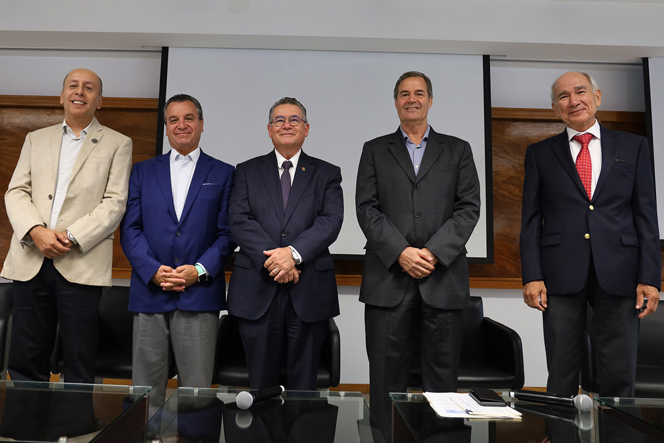
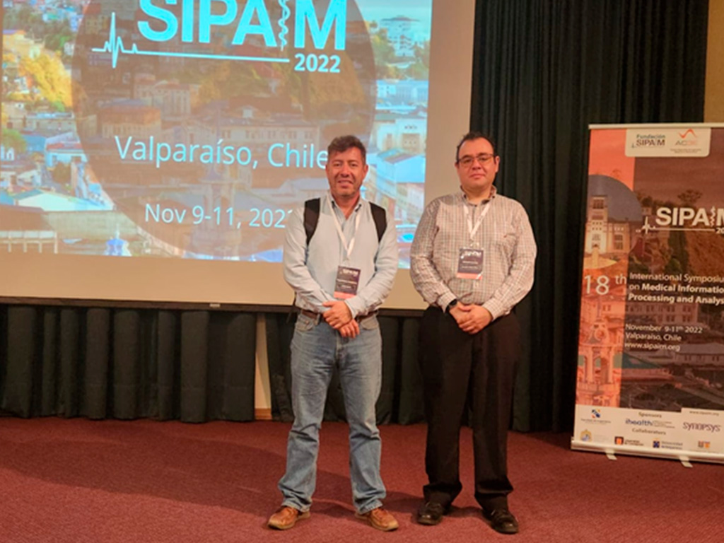
Between November 9 and 11, 2022, Dr. Ernesto Moya-Albor and Dr. Jorge Brieva, research professors from the School of Engineering, Mexico City campus, attended the 18th International Symposium on Medical Information Processing and Analysis (SIPAIM 2022) held in Valparaiso, Chile.
SIPAIM 2022
In this edition of SIPAIM, both researchers, together with faculty research professor Dr. Karina Ruby Pérez Daniel and in collaboration with scholars from the National Polytechnic Institute and the Center for Research in Mathematics, presented two research papers.
Both Dr. Brieva, who is a SIPAIM Steering Committee member, and Dr. Moya-Albor were part of the Scientific Committee. Dr. Brieva also participated as moderator of plenary lectures and on the event’s organizational committee.
Modeling of the Major Temporal Arcade
With their article “Modeling of the Major Temporal Arcade,” using genetic algorithms and orthogonal polynomials, the researchers presented a novel method of verifying the state of the retina and detecting changes in the vascular structure, both in adults and in children with diabetic retinopathy, one of the main causes of blindness.
For this purpose, the researchers presented a numerical model of the major temporal arcade in funduscopy images using orthogonal Legendre, Chebyshev, and Laguerre polynomials and an evolutionary algorithm. A set of twenty funduscopy images was used to evaluate the proposal, where an expert performed the delineation of the major temporal arch.
The proposed method was compared with three other state-of-the-art methods, and the study found that Legendre polynomials are the most suitable for generating a tool to aid in computer-assisted ophthalmologic diagnosis.
The authentication of medical images
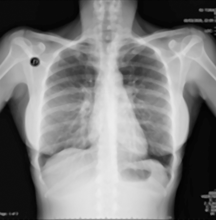
Similarly, the researchers participated in the presentation entitled, “The Authentication of Medical Images through a Hybrid Watermarking Method based on Hermite-Jigsaw-SVD,” where they discussed results from a robust method for watermarking medical images.
The doctors used the Steered Hermite transform (SHT), a spatial and frequency image decomposition technique inspired by the human vision system, to invisibly embed the watermark.
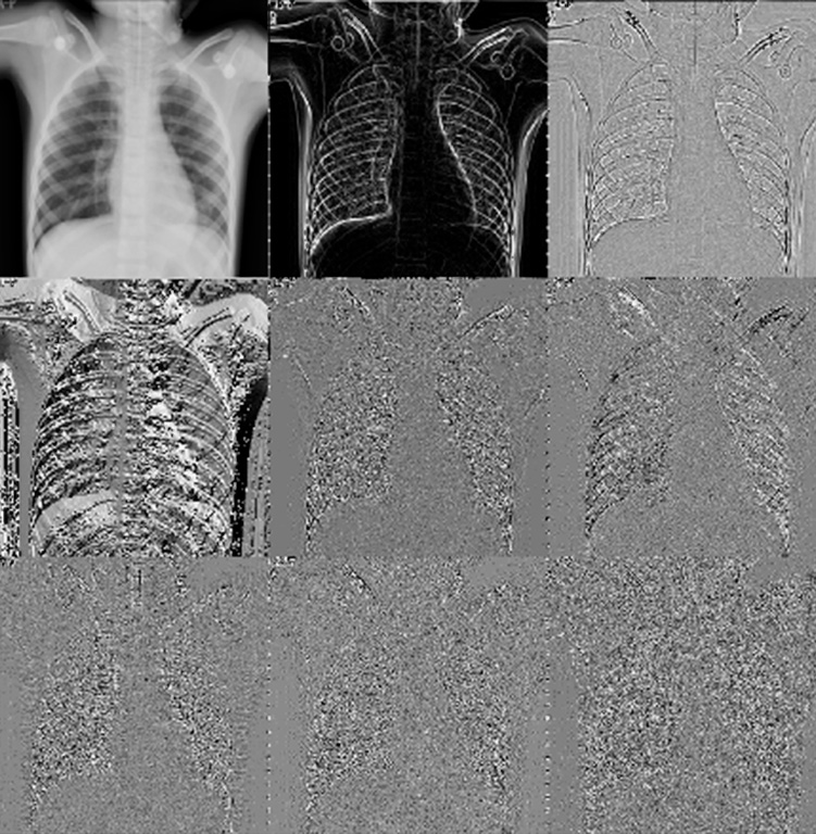
They also included singular value decomposition (SVD) in the proposal to obtain greater robustness of watermarks against attacks. They also used the Jigsaw transform to encrypt watermarks before embedding them in medical images. Finally, they used 40 X-ray images and the caduceus medical symbol as a watermark to evaluate the proposed watermarking method.
The results showed that the watermark is invisible to the human eye and is extracted without any loss of visual information. Furthermore, the marked medical images do not appear to have any significant visual alterations, preserving visual information for the patient’s diagnosis.
Furthermore, in tests involving attacks on a marked medical image, the results verified that the proposed method is robust and can stand up against common geometric and processing attacks (Gaussian noise, salt and pepper noise, median filter, JPEG compression, rotation, and translation), allowing the perceptible extraction of watermarks even in strong attacks where the marked medical image has lost a lot of information.


